Introduction
The buccal cavity is formed by the cheeks, the hard and soft palates (the roof of the mouth) and tongue. The cheeks form the lateral walls of the buccal cavity, which are covered by non-keratinised stratified squamous epithelium. The hard palate, which is formed by the roof bones, is covered by mucous membrane and forms a bony partition between the buccal cavity and the nasal cavity. The soft palate forms the back (muscular) portion of the roof and is also lined by mucous membrane (Christie et al., 1995).
Oral mucosa
The thickness of the buccal mucosa does not vary to a great extent in different species. The mucosa of rat is keratinised which makes this species unsuitable for research on buccal drug delivery. More useful models are the rabbit and dog buccal mucosa both of which have been claimed to be broadly similar in structure and composition to human buccal mucosa species (Harris and Robinson, 1992).
The pH membrane permeability relationship in the mucosa observed for humans was also observed in dogs (Barsuhn et al., 1988). Active transport, indicated by measurement of the potential difference across the mucosa, was also found in dogs and rabbits. The observed potential difference across the buccal mucosa of humans, dogs and rabbits differed slightly (Utoguchi et al., 1997). However, a correlation between similar potential difference over the mucosa and a similar active transport is unclear.
Saliva and salivary glands
Saliva can influence the micro-flora in the buccal cavity. For instance parotid proteins (high molecular weight glycoproteins), submandibular and sublingual mucin induce aggregations of oral bacteria strains in humans (Koop et al., 1990), which are also observed in other animals such as streptococcus and actinomyces viscous in rats (de Jong et al., 1984). Although little information is available, it is not inconceivable that microflora in the buccal cavity of the different animal species can differ to a great extent.
In humans the three major salivary glands are the parotid, submandibular and sublingual glands. They are also found in mouse, rat, rabbit, dog, monkey and pig. Minor salivary glands in humans are buccal, palatal and lingual glands. Some of these minor salivary glands have been observed in mouse, rat, rabbit, dog, monkey and pig. The rabbit sublingual gland should be named actually the minor sublingual gland, as the major sublingual gland seen in many mammals is not present. On the other hand a zygomatic salivary gland is present in rabbit, which is not present in human and other species (Manning et al., 1994).
The human mouth
In human saliva is mainly produced by the greater salivary glands (parotid gland 23%, submandibular gland 65%, sublingual gland 4%). The minor salivary glands produce only 8% of the total volume of saliva Hold et al., 1998).
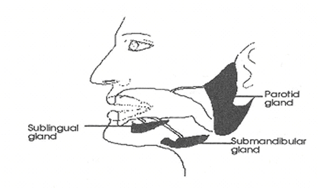
Figure 1: Position of the human salivary glands (Wilson and Washington, 1989,p24)